A vitrectomy is a type of eye surgery to treat problems with the retina and vitreous. During the surgery, your surgeon removes the vitreous and replaces it with another solution.
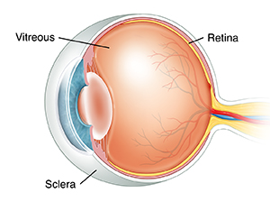
What are the retina and vitreous?
The retina is a layer of cells at the back of your eye. These cells use light to send visual information to your brain. The vitreous is a gel-like substance that fills the eye. Normally the vitreous is clear. This is so light can pass through the eye and reach the retina. But some problems can cause blood and debris to block the light. The vitreous can pull or tug on your retina, causing a retinal hole or tear. This can harm your vision.
Why vitrectomy is done
A vitrectomy is 1 type of treatment for any of these eye problems:
-
Diabetic retinopathy
-
Detached retina
-
Vitreous hemorrhage
-
Infection inside your eye
-
Severe eye injury
-
A hole in the central part of your retina (macula)
-
A wrinkle in the central part of your retina
-
Certain problems after cataract surgery
All of these problems can cause vision loss. If not treated, some of them can even lead to blindness. Vitrectomy can often restore lost vision. You might need a vitrectomy done as an emergency if you have an eye injury. In other cases, your eye healthcare provider will schedule your vitrectomy in advance. Vitrectomy is sometimes done for a detached retina. Removing the vitreous gives better access to your retina and decreases the tension on your retina.
How vitrectomy is done
You may have general anesthesia to put you to sleep. If this is the case, you will sleep through the surgery. Or you may be sedated but awake during the surgery. You will get medicine to help you relax. You may also be given anesthetic eye drops and injections. These are to numb the eye and make sure that you don’t feel anything during the surgery.
A small cut is made in the white part of the eye (sclera). The gel-like vitreous is removed, along with any scar tissue or other material. Repairs are made to the eye as needed. The vitreous is then replaced with another type of fluid. It may be replaced with silicone oil or saltwater (saline) solution. Or it may be replaced by air or gas. This will depend on the problem that is being treated.
Risks of vitrectomy
All surgery has some risks. The risks of vitrectomy include:
-
Infection
-
Bleeding
-
High pressure in the eye
-
New retinal detachment caused by the surgery
-
Damage to the lens
-
Increased rate of cataract formation
-
Problems with eyesight, such as double vision
-
Change in refractive error
-
Repair is not successful
-
Need for a repeat surgery